Contaminant identification in many varied industries requires the ability to pinpoint single microscopic particles and subsequently identify and/or characterise them.
Micro-XRF is the ideal tool to analyse these contaminant particles. The penetrating nature of the primary X-ray beam allows particles which are below the surface to be easily analysed – and even particles which are invisible to the eye can be located and identified using XRF and transmitted X-ray imaging. Beam sizes ranging from 10 µm through to 1.2 mm ensure that both micro and macro contaminants can be analysed easily and quickly.
The sub-surface analysis capability of micro-XRF is often a key point in making this technique the first point of call for scientists. Compared with SEM/EDX analysis, which has traditionally been used in this field, micro-XRF offers a considerable gain in ease of use, and is suitable for the analysis of large areas of real life samples without the need for sample cutting and preparation. Unlike SEM/EDX, micro-XRF systems can be operated without vacuum, thus allowing easy analysis of biological and food materials.
SLICE software offers a comprehensive data archiving tool for micro-XRF and SEM/EDX data, and is an invaluable tool for analysts who encounter varied contaminant materials during their work. In addition, the database with many thousands of spectra of common metals and glasses is key in providing a fast positive identification of the analysed contaminant.
- Non-destructive identification of microscopic contaminant particles
- Location of foreign objects at and below the sample surface
- Analysis of large real life samples for
- Electronics
- Engine wear debris
- Food and beverage
- Manufacturing
- Pathology
- Pharmaceutics
- Semiconductor
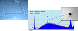
A microscopic foreign particle observed in a molded plastic dish is pinpointed using the XGT’s 100 µm X-ray beam. The XRF spectrum of the particle provides a positive identification of zinc, originating from the machinery used within the factory.

Phosphorus is often added to integrated chip molds as a fire retardant. Reaction with water vapour can result in phosphoric acid which then melts the copper circuitry. In these images, chip failure points invisible to the eye have been observed using transmitted X-ray imaging. Detailed XRF imaging highlights the presence of anomalous Phosphorus hotspots, and subsequent copper ion migration caused by melting.
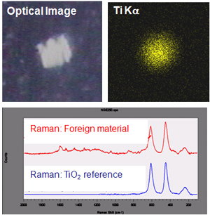
A white particle in a factory produced resin is identified as a titanium species using micro-XRF. Complementary analysis using a LabRAM Raman microscope positively identifies the material as being TiO2.
